


Development and Characterization of a New Cell Line Derived from the Kidney of Large Yellow Croaker(Larimichthys crocea)
-
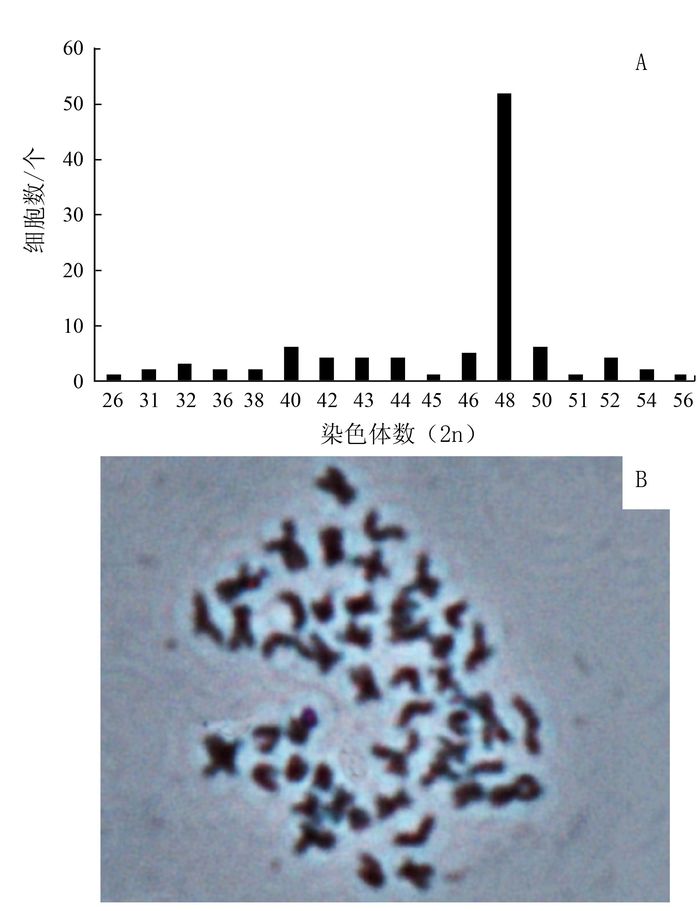
摘要: 大黄鱼Larimichthyscrocea是福建省最重要的经济性鱼类之一。为扩充完善大黄鱼体细胞库,丰富其病毒学、细胞毒理学及细胞工程学研究手段,以大黄鱼肾脏组织为材料,进行组织细胞体外培养条件的探索,构建大黄鱼体细胞体外培养技术体系,并在此基础上建立大黄鱼肾组织细胞系(YCK)。原代培养使用组织块贴壁翻瓶法,标准传代培养是以0.25%胰蛋白酶消化细胞,使用添加10%胎牛血清(FBS)的M-199,在27℃下进行密闭培养,每72 h完全换液1次。该细胞系主要由上皮样细胞构成,在上述培养条件下生长旺盛,经过400多天的连续培养,已经成功传代超过70次,第65代YCK细胞的群体倍增时间为36.12 h。对常用的鱼类细胞培养基进行适应性筛选,确认M-199是最适合该细胞系的培养介质,也可适用于其他大黄鱼细胞的培养。对该细胞系的染色体分析表明:虽然出现了异倍化现象,但该系依然符合2n=48的二倍体细胞系。Abstract: Large yellow croaker (Larimichthys crocea) is an important economic species in Fujian. To develop virological, toxicological and cell engineering techniques, as well as resources for advanced researches on the fish, this study had developed a new cell line derived from the fish kidneys(YCK). The obtained YCK has been successfully conserved in the M-199 medium supplemented with 10% fetal bovine serum at 27℃ over 400 days with a medium-change every 72 h, and satisfactorily sub-cultured more than 70 times during the interim. A parallel test of commonly applied culture media was conducted to get the conclusion that M-199 being the most suitable medium for YCK. This newly established cell line consisted predominantly of epithelial-like cells, and was proven to be diploid with a modal chromosome number of 2n=48.
-
Key words:
- large yellow croaker (Larimichthys crocea) /
- kidney /
- cell line
-

图 1 大黄鱼肾组织细胞的原代和传代培养
注:A为大黄鱼肾脏原代培养,第12 d;B为第6代YCK细胞(标尺100 μm);C为第27代YCK细胞;D为第70代YCK细胞。
Figure 1. Primary culture and subculture of cells from kidney of L. crocea
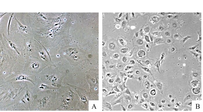
图 2 大黄鱼肾组织细胞系YCK在不同培养基的生长情况
注:A为L-15中培养的YCK细胞;B为RPMI-1640中培养的YCK细胞。
Figure 2. Comparison of the growth characteristics of YCK in different culture media
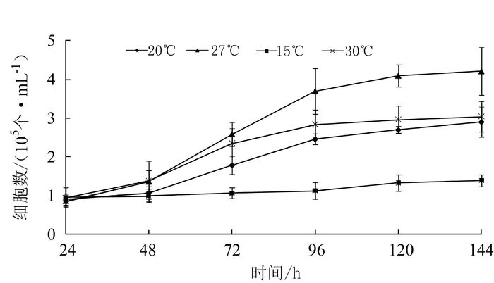
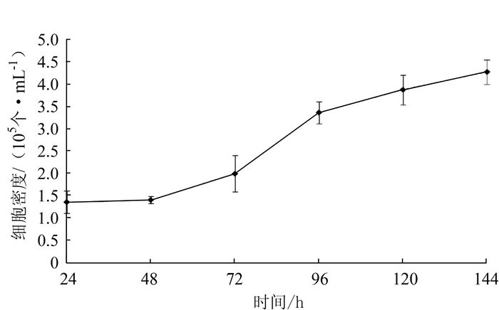
图 3 不同培养温度下大黄鱼肾组织细胞系YCK生长趋势
Figure 3. Growth kinetics of YCK under different temperatures
-
[1] WOLF K, QUIMBY M C. Established eurythermic line of fish cells in vitro[J]. Science, 1962, 135(3508):1065-1066. doi: 10.1126/science.135.3508.1065 [2] FRYER J L, LANNAN C N. Three decades of fish cell culture:A current listing of cell lines derived from fish[J]. Journal of Tissue Culture Methods, 1994, 16(2):87-94. doi: 10.1007/BF01404816 [3] BOLS N C, LEE L E J.Technology and uses of cell cultures from the tissuesand organs of bony fish[J].Cytotechnology, 1991, 6(3):163-187. doi: 10.1007/BF00624756 [4] CHEN S N, KOU G H. Establishment, characterization and application of 14 cell lines from warm water fish[C]//Karoda Y, Kursta E, Maramorosch K, et al.Invertebrate and Fish Tissue Culture, Germany:Springer-Verlag, Berlin, 1988:218-227. [5] BAKSI S M, FRAZIER, J M. Isolated fish hepatocytes model systems for toxicology research[J]. Aquatic Toxicology, 1990, 16(4):229-256. doi: 10.1016/0166-445X(90)90039-R [6] ZHANG Q Y, RUAN H M, LI Z Q, et al. Infection and propagation of Lymphocystis virus isolated from the cultured flounder Paralichthys olivaceus in grass carp cell lines[J]. Diseases of Aquatic Organisms, 2003, 57(1-2):27-34. https://www.sciencedirect.com/science/article/pii/S1050464817307076 [7] 张奇亚, 桂建芳.水生病毒学[M].北京:高等教育出版社, 2007:35-50. [8] 樊海平.福建省水生动物病害防控学科发展现状与设想[J].福建水产, 2011, 33(3):48-50. http://www.cqvip.com/QK/94635X/201103/39584200.html [9] 王小文, 陈新华.大黄鱼虹彩病毒腺苷三磷酸酶(ATPase)基因的克隆与表达[J].病毒学报, 2004, 20(1):81-85. http://kns.cnki.net/KCMS/detail/detail.aspx?filename=bdxb200401015&dbname=CJFD&dbcode=CJFQ [10] 孙爱. 大黄鱼三种组织细胞系的建立、鉴定及其应用的初步研究[D]. 青岛: 中国海洋大学, 2010. http://www.doc88.com/p-8826242151844.html [11] DONG C, WENG S, SHI X, et al. Development of a mandarin fish Siniperca chuatsifry cell line suitable for the study of infectious spleen and kidney necrosis virus (ISKNV)[J]. Virus Research, 2008, 135(2):273-281. doi: 10.1016/j.virusres.2008.04.004 [12] DAVIS J M. Basic Cell Culture, 2nd Edition[M]. U K:Oxford University Express Oxford, 2001:311-405. [13] LEVAN A, FREDGA K, SANDBERG A.Nomenclature for centromeric position on chromosomes[J]. Hereditas, 1964, 52(2):201-220. doi: 10.1007/BF00938015 [14] LAKRA W S, RAJA S T, JOY K P. Development, characterization, conservation and storage of fish cell lines:a review[J]. Fish Physiology and Biochemistry, 2011, 37(1):1-20. doi: 10.1007/s10695-010-9411-x [15] 董传甫. 斑石鲷虹彩病毒(SKIV)结构蛋白质组学及三个新型鱼类细胞系的建立与部分特性[D]. 广州: 中山大学, 2010. http://d.wanfangdata.com.cn/Thesis/Y1779289 [16] 樊廷俊, 耿晓芬, 丛日山, 等.大菱鲆鳍细胞系的建立[J].中国海洋大学学报, 2007, 37(5):759-766. http://d.wanfangdata.com.cn/Thesis/Y1112077 [17] 张博, 陈松林.近10年鱼类细胞培养研究进展及应用展望[J].海洋科学, 2011, 35(7):113-121. http://www.cqvip.com/QK/90010X/201107/38801144.html [18] WOLF K, QUIMBY M C. Procedures for subculturing fish cells and propagating fish cell lines[J]. Tissue Culture Association Manual, 1976, 2(4):471-474. doi: 10.1007/BF00918344 [19] BRYSON S P, JOYCE E M, MARTELL D J, et al. A cell line (HEW) from embryos of haddock(Melanogrammus aeglefinius) and its capacity to tolerate environmental extremes[J]. Marine Biotechnology, 2006, 8(6):641-653. doi: 10.1007/s10126-005-6163-1 [20] 张彩兰, 刘家富, 李雅璀, 等.福建省大黄鱼养殖现状分析与对策[J].上海水产大学学报, 2002, 11(1):77-84. http://www.oalib.com/paper/5105887 [21] LANNAN C N. Fish cell culture:A protocol for quality control[J]. Journal of Tissue Culture Methods, 1994, 16(2):95-98. doi: 10.1007/BF01404817 [22] LEIBOVITZ A. The growth and maintenance of tissue-cell cultures in free gas exchange with the atmosphere[J]. American Journal of Hygiene, 1963, 78(2):173-180. https://www.cabdirect.org/cabdirect/abstract/19642701158 [23] LEIBOVITZ A. Preparation of medium L-15[J]. Tissue Culture Association Manual, 1977, 3(2):557-559. doi: 10.1007/BF00918380 [24] YU H, COOK T J, SINKO P J. Evidence for diminished functional expression of intestinal transporters in Caco-2 cell monolayers at high passages[J].Pharmacological Research, 1997, 14(6):757-762. doi: 10.1023/A:1012150405949 [25] DOYLE A, GRIFFITHS J B. Cell and tissue culture for medical research[M]. UK:John Wiley & Sons Ltd, West Sussex, 2000:102-128. [26] ZHOU L R, DEANE E E, WOO N Y S. Development of a black sea bream fibroblast cell line and its potential use as an in vitro model for stress protein studies[J]. Fish Physiology and Biochemistry, 2003, 29:255-262. http://www.nature.com/cited/cited.html?doi=10.1038/emm.2002.19 [27] 王德祥, 苏永全, 王世锋.不同地理种群大黄鱼染色体核型的比较研究[J].海洋学报, 2006, 28(16):176-179. http://www.wenkuxiazai.com/doc/ae44fbb269dc5022aaea00c5.html [28] 樊廷俊, 魏云波, 徐晓辉.褐点石斑鱼三种组织细胞系的建立[J].中国海洋大学学报, 2009, 39(5):961-964. http://www.cnki.com.cn/Article/CJFDTotal-QDHY200905031.htm [29] SWAMINATHAN T R, KUMAR R, JENCY P M E, et al.A new fish cell line derived from the caudal fin of freshwater angelfish Pterophyllum scalare:development and characterization[J].Journal of Fish Biology, 2016, 89(3):1769-1781. doi: 10.1111/jfb.2016.89.issue-3 [30] ZHENG Y, WANG N, XIE M S, et al. Establishment and characterization of a new fish cell line from head kidney of half-smooth tongue sole (Cynoglossus semilaevis)[J].Fish Physiology and Biochemistry, 2012, 38(6):1635-1643. doi: 10.1007/s10695-012-9660-y -

 下载:
下载:
 点击查看大图
计量
- 文章访问数: 1630
- HTML全文浏览量: 259
- PDF下载量: 121
- 被引次数: 0
