


SYBR Green I-based RT-PCR Assay for Detecting IFN-β mRNA in Duck
-
摘要:
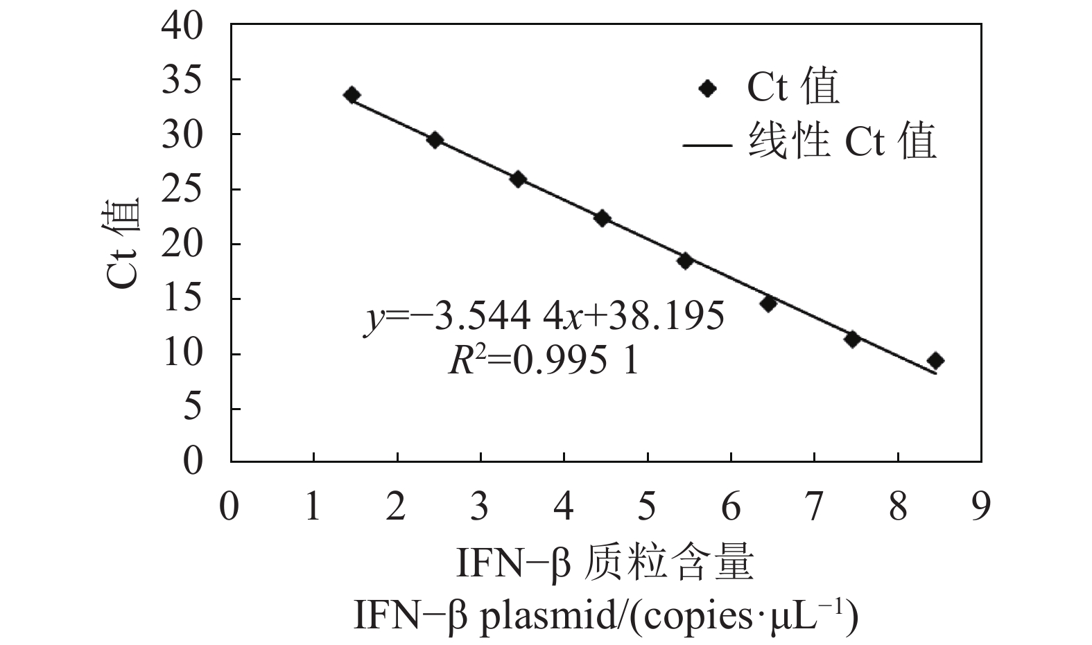
目的 建立一种检测鸭IFN-β mRNA转录水平的SYBR Green Ⅰ实时荧光定量RT-PCR检测方法。 方法 根据GenBank中鸭IFN-β(KT428159)核苷酸序列设计并合成特异性引物,将鸭IFN-β基因克隆至pET-30a载体,以此构建的pET-30a-IFN-β阳性重组质粒作为阳性标准品,采用SYBR Green Ⅰ实时荧光定量PCR检测,构建标准曲线,并进行引物特异性、灵敏度及重复性试验。 结果 该扩增特异性强,无引物二聚体及非特异性产物,熔解曲线单峰(Tm=87.94±0.16 ℃);Ct值在8.9~34.0线性拟合程度高,相关系数R2>99.5%;灵敏度高,最低检测限为2.84 copies·μL−1;重复性好,对来自临床的3种组织样品检测的组内变异系数小于0.13%,组间变异系数不超过1%。 结论 该方法特异性强、灵敏度高、重复性好,为鸭IFN-β mRNA表达水平的定量分析提供了技术手段。 -
关键词:
- 鸭 /
- IFN-β /
- 荧光定量RT-PCR /
- SYBR Green I /
- mRNA
Abstract:Objective A method for detecting IFN-β in duck using SYBR Green I-based RT-PCR was developed. Methods A pair of specific primers was designed according to the GenBank nucleotide sequence on IFN-β of duck (KT428159). The gene was cloned into a pET-30a vector, and the recombinant plasmid pET-30a-IFN-β severed to establish a standard curve. The specificity, sensitivity, and repeatability of the new methodology were determined. Result The melting curves of measurement showed a sharp single peak at Tm=87.94±0.16 ℃ without non-specific amplification and primer dimers, indicating high specificity of the methodology. The Ct value ranged from 8.9 to 34.0 with a standard curve showing a linearity with R2>99.5%. The detection limit on IFN-β was 2.84 copies/μL. The repeatability on the Ct data for the intra- and inter-groups had coefficients of variation below 0.13% and 1%, respectively. Conclusion The newly developed assay was specific, sensitive, repeatable, and suitable for the quantitative detection of IFN-β mRNA in ducks. -
Key words:
- Duck /
- IFN-β /
- qRT-PCR /
- SYBR Green I /
- mRNA
-
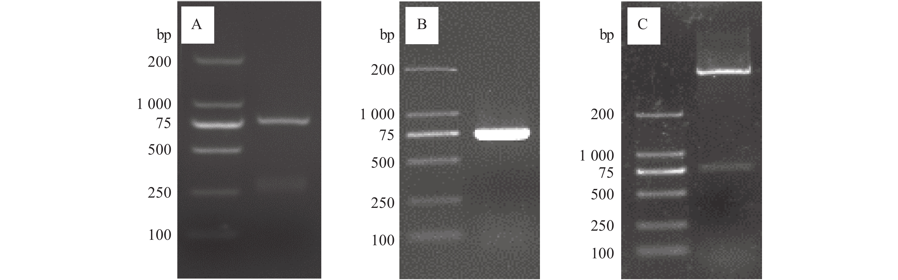
图 1 鸭IFN-β全基因的PCR扩增及重组质粒的构建
注:A,鸭脾脏IFN-β基因的扩增;B,重组质粒的IFN-β基因扩增;C,重组质粒的双酶切鉴定。
Figure 1. PCR amplification of duck IFN-β and construction of recombinant plasmid
Note: A: amplification of IFN-β from duck spleen; B: IFN-β amplification from recombinant plasmid; C: identification of recombinant plasmid by double restriction enzyme digestion.
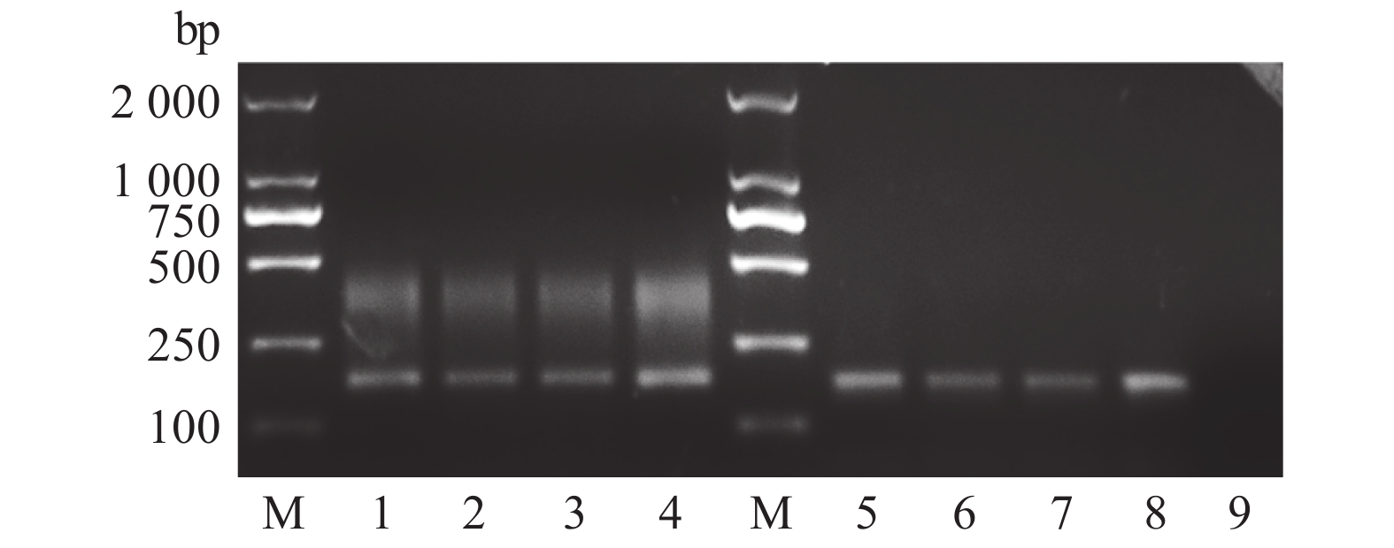
图 2 不同组织IFN-β的常规RT-PCR和荧光定量RT-PCR
注:1~4分别为脾脏、肝脏、胰腺及DEFs细胞的常规PCR扩增;M为DL 2000 DNA Marker;5~8分别为脾脏、肝脏、胰腺及DEFs细胞的qPCR扩增;9、DEPC水。
Figure 2. Conventional RT-PCR and qRT-PCR amplification of IFN-β in different tissues
Note: 1–4: conventional PCR amplification of IFN-β in spleen, liver, pancreas, and DEFs cells, respectively; M: DL 2000 DNA marker; 5–8: qPCR amplification of IFN-β in spleen, liver, pancreas, and DEFs cells, respectively; 9: DEPC water.
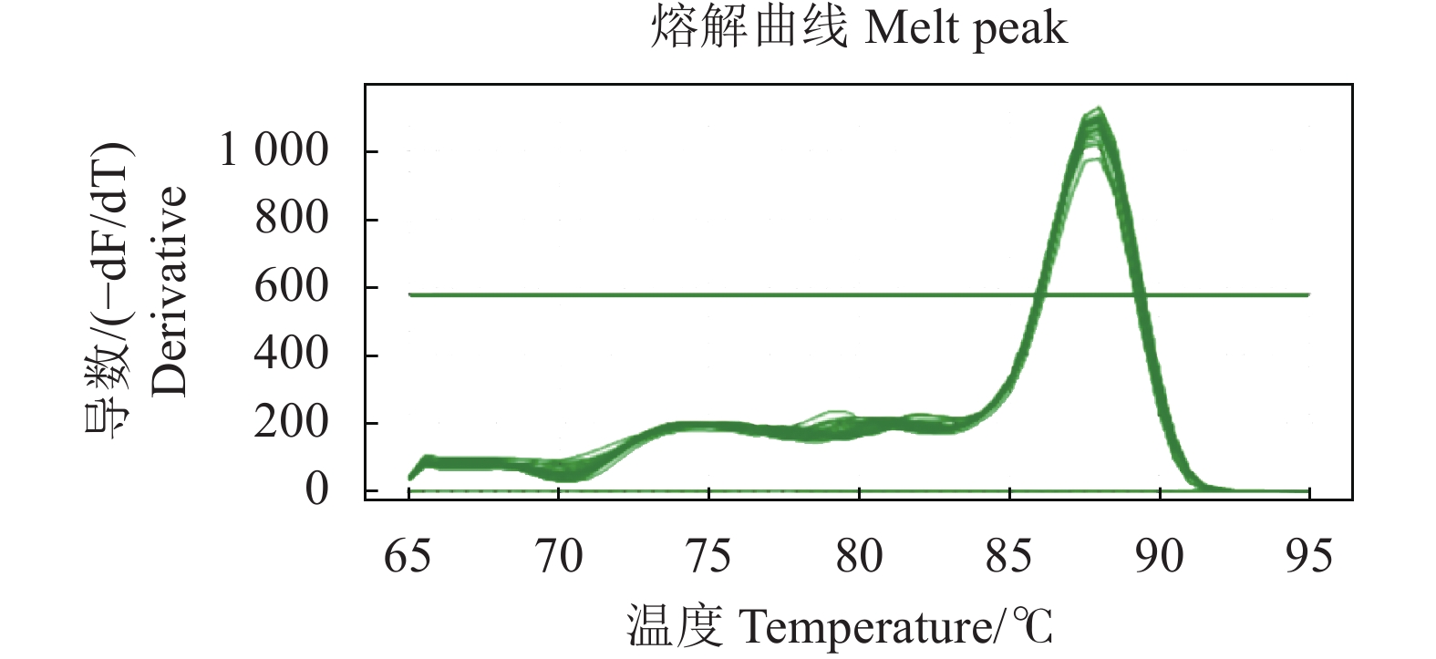
图 3 鸭4种组织(肝、脾、胰和DEFs)IFN-β 荧光定量RT-PCR熔解曲线
Figure 3. qRT-PCR melting curve of duck IFN-β
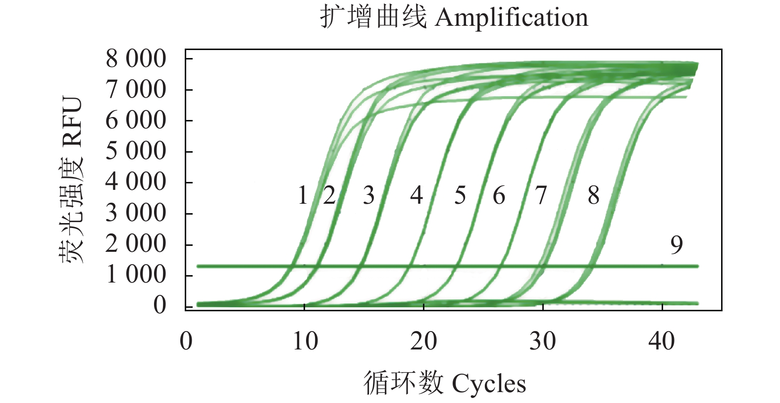
图 4 IFN-β 荧光定量RT-PCR扩增曲线
注:1~8分别为2.84×10⁸、2.84×10⁷、2.84×10⁶、2.84×10⁵、2.84×10⁴、2.84×10³、2.84×10²、2.84×101 copies·μL−1;9、DEPC水。
Figure 4. Amplification curve of qRT-PCR for IFN-β
Note: 1–8: 2.84×10⁸, 2.84×10⁷, 2.84×10⁶, 2.84×10⁵, 2.84×10⁴, 2.84×10³, 2.84×10², and 2.84×101 copies·μL−1, respectively. 9: DEPC water.
表 1 引物核苷酸序列及扩增片段大小
Table 1. Primer sequences and amplified fragment sizes
引物
Primers核苷酸序列
Nucleotide sequence(5′-3′)扩增片段大小
Amplified fragment size/bpFull-IFN-β-F CGCGGATCCATGCCTGGGCCATCAGC 729 Full-IFN-β-R CCCAAGCTTTCACGCCGTGGGCTTGT qIFN-β-F GGGCTCCGCAACCTTCACC 165 qIFN-β-R TGCTTGGCTCTTCATCCGCCGTA 注:下划线区域为酶切位点序列。
Note:The underlined parts are the sequences of restriction sites. 下载: 导出CSV
表 2 实时荧光定量RT-PCR敏感性检测
Table 2. Sensitivity of qRT-PCR assay
质粒拷贝数
Copy number
/(Copies·μL−1)2.84×103 2.84×10² 2.84×101 2.84×100 2.84×10⁻¹ Ct值(平均值±标准差)
Ct (Means±SD)26.38±0.03 29.87±0.10 33.98±0.05 36.13±1.12 − 变异系数 CV/% 0.11% 0.33% 0.15% 3.10% −
下载: 导出CSV
表 3 实时荧光定量PCR方法的批内与批间重复性评价结果
Table 3. Reproducibility of intra- and inter-qRT-PCR assays
组织
Tissue组内重复性试验Ct值
The Ct values of intra-assay组间重复性试验Ct值
The Ct values of inter-assay平均值±标准差
Means±SD变异系数
CV/%平均值±标准差
Means±SD变异系数
CV/%脾 Spleen 26.13±0.03 0.11 26.37±0.21 0.80 肝 Liver 32.63±0.04 0.12 32.41±0.20 0.62 胰 Pancreas 31.51±0.04 0.13 31.54±0.30 0.95
下载: 导出CSV
-
[1] WALTER M R. The role of structure in the biology of interferon signaling [J]. Frontiers in Immunology, 2020, 11: 606489. doi: 10.3389/fimmu.2020.606489 [2] RAI K R, SHRESTHA P, YANG B C, et al. Acute infection of viral pathogens and their innate immune escape [J]. Frontiers in Microbiology, 2021, 12: 672026. doi: 10.3389/fmicb.2021.672026 [3] 万春和, 朱海侠, 陈红梅, 等. 番鸭IFN-α mRNA实时荧光定量RT-PCR检测方法的建立 [J]. 中国动物传染病学报, 2012, 20(4):63−68. doi: 10.3969/j.issn.1674-6422.2012.04.012WAN C H, ZHU H X, CHEN H M, et al. Development of a quantitative real-time PCR for detection of IFN-α mRNA of Muscovy ducks [J]. Chinese Journal of Animal Infectious Diseases, 2012, 20(4): 63−68.(in Chinese) doi: 10.3969/j.issn.1674-6422.2012.04.012 [4] 刘澜澜, 庄艳娜, 于晓红, 等. 绿头鸭IFN-α的可溶性表达及其活性分析 [J]. 生物技术通报, 2014(11):142−146.LIU L L, ZHUANG Y N, YU X H, et al. Soluble expression and activity analysis of mallard IFN-Α [J]. Biotechnology Bulletin, 2014(11): 142−146.(in Chinese) [5] 杨发龙, 谢秀兰, 汤承, 等. 鸭瘟病毒疫苗株与强毒株诱导雏鸭IFN-α mRNA在肝脏中表达的动态定量研究 [J]. 中国预防兽医学报, 2008, 30(8):647−650.YANG F L, XIE X L, TANG C, et al. Dynamics of IFN-α mRNA expression in liver of ducks infected with duck plague virus of different virulence [J]. Chinese Journal of Preventive Veterinary Medicine, 2008, 30(8): 647−650.(in Chinese) [6] 高全新, 刘云霞, 程玉强, 等. 鸭IFN-β启动子双荧光素酶报告基因系统的构建及活性检测 [J]. 上海农业学报, 2018, 34(3):66−71.GAO Q X, LIU Y X, CHENG Y Q, et al. Construction and activity detection of dual luciferase reporter gene system for duck IFN-β promoter [J]. Acta Agriculturae Shanghai, 2018, 34(3): 66−71.(in Chinese) [7] CHEN Z L, LUO G F, WANG Q X, et al. Muscovy duck reovirus infection rapidly activates host innate immune signaling and induces an effective antiviral immune response involving critical interferons [J]. Veterinary Microbiology, 2015, 175(2/3/4): 232−243. [8] LI N, WANG Y, LI R, et al. Immune responses of ducks infected with duck Tembusu virus [J]. Frontiers in Microbiology, 2015, 6: 425. [9] 张盼涛, 曾显营, 杨婧, 等. 鸭IFN-γ, IL-2 mRNA荧光定量RT-PCR方法的建立及应用 [J]. 中国预防兽医学报, 2013, 35(6):472−476. doi: 10.3969/j.issn.1008-0589.2013.06.11ZHANG P T, ZENG X Y, YANG J, et al. Establishment and application of a real-time assay for detecting of IFN-γ and IL-2 mRNA in ducks [J]. Chinese Journal of Preventive Veterinary Medicine, 2013, 35(6): 472−476.(in Chinese) doi: 10.3969/j.issn.1008-0589.2013.06.11 [10] PIZZATO M, ERLWEIN O, BONSALL D, et al. A one-step SYBR Green I-based product-enhanced reverse transcriptase assay for the quantitation of retroviruses in cell culture supernatants [J]. Journal of Virological Methods, 2009, 156(1/2): 1−7. [11] TAKEUCHI O, AKIRA S. Innate immunity to virus infection [J]. Immunological Reviews, 2009, 227(1): 75−86. doi: 10.1111/j.1600-065X.2008.00737.x [12] 陈超, 池晓娟, 白庆玲, 等. 甲型流感病毒感染过程中干扰素介导的天然免疫应答机制 [J]. 生物工程学报, 2015, 31(12):1671−1681.CHEN C, CHI X J, BAI Q L, et al. Mechanisms underlying interferon-mediated host innate immunity during influenza A virus infection [J]. Chinese Journal of Biotechnology, 2015, 31(12): 1671−1681.(in Chinese) [13] LI N, HONG T Q, LI R, et al. Cherry valley ducks mitochondrial antiviral-signaling protein-mediated signaling pathway and antiviral activity research [J]. Frontiers in Immunology, 2016, 7: 377. [14] ZHANG H H, SONG X D, LI T X, et al. DDX1 from Cherry valley duck mediates signaling pathways and anti-NDRV activity [J]. Veterinary Research, 2021, 52(1): 9. doi: 10.1186/s13567-020-00889-4 [15] LI N, JIANG S N, ZHAO J, et al. Molecular identification of duck DDX3X and its potential role in response to Tembusu virus [J]. Developmental and Comparative Immunology, 2020, 106: 103599. doi: 10.1016/j.dci.2019.103599 [16] HE T Q, WANG M S, CHENG A C, et al. Duck enteritis virus pUL47, as a late structural protein localized in the nucleus, mainly depends on residues 40 to 50 and 768 to 777 and inhibits IFN-β signalling by interacting with STAT1 [J]. Veterinary Research, 2020, 51(1): 1−12. doi: 10.1186/s13567-019-0731-2 [17] ZHANG W, JIANG B W, ZENG M, et al. Binding of duck tembusu virus nonstructural protein 2A to duck STING disrupts induction of its signal transduction cascade to inhibit beta interferon induction[J]. Journal of Virology, 2020, 94(9). DOI: 10.1128/jvi.01850-19. -

 点击查看大图
计量
- 文章访问数: 545
- HTML全文浏览量: 118
- PDF下载量: 23
- 被引次数: 0
