


Genomic Sequencing and VP1 Polyclonal Antibody Preparation for a Duck Hepatitis Virus
-
摘要:
目的 确定福建某鸭场雏鸭以出现角弓反张和肝脏出现肿大并伴有密集出血点为主要病征的高死亡率病害的病原,并制备可特异性检测的多克隆抗体,以期为福建省鸭流行病学研究提供素材。 方法 取该鸭场病死鸭肝脏组织进行病毒的分离鉴定,并对病毒进行全基因测序。利用pGEX-4T-1载体构建该病毒优势抗原表位基因的高效表达系统,制备VP1多克隆抗体,并用Western-blotting检测其特异性。 结果 应用非免疫的鸭胚从疑似鸭病毒性肝炎的鸭肝脏中分离到1株病毒。该分离毒在鸭胚连续传6代后死亡率约为70%,且死胚尿囊液分别在鸭、鸡、小鼠和兔血液中均未出现血凝现象。注射分离毒的尿囊液的1日龄雏鸭死亡率达100%。RT- PCR扩增结果、全基因组测序结果表明分离毒为I型鸭病毒性肝炎病毒,毒株DQ226541.1的基因序列同源性达99.4%,其VP1氨基酸序列与毒株DQ226541.1氨基酸序列一致。将毒株命名为Fujian2015。成功构建重组质粒pGEX-4T-1-VP1,并制备了可特异性检测VP1蛋白的VP1多抗血清。 结论 成功分离到了1株I型鸭病毒性肝炎病毒,同时制备了该毒株的VP1多克隆抗体,为后续检测I型鸭病毒性肝炎病毒相关研究奠定了基础。 Abstract:Objective The pathogen that caused the high mortality disease showing symptoms of anterior arch reflexion and liver enlargement with dense bleeding points on ducklings at a duck farm in Fujian was identified. Specifically detectable polyclonal antibody was prepared for epidemiological study on the disease. Method Liver tissues of the affected and died ducklings were collected for virus identification. Sequence of the virus was determined, and the pGEX-4T-1 vector used to construct a high-efficiency expression system for the dominant epitope gene. A VP1 polyclonal antibody was prepared, and specificity confirmed by western-blotting. Result A virus was isolated from the liver of a duck suspected of viral hepatitis using non-immune duck embryos. The mortality rate by the isolated virus on the embryos after 6 generations of continuous transmission was approximately 70%. But the allantoic fluid of the dead embryo did not show hemagglutination in the blood of duck, chicken, mouse, or rabbit. In contrast, the mortality rate on 1-day-old ducklings injected with the allantoic fluid was 100%. The RT-PCR amplification and the entire genome sequence indicated the isolated virus to be positive for duck viral hepatitis virus 1. Furthermore, the gene homology between the virus and DQ226541.1 was 99.4%, and the sequence of VP1 amino acids consistent with that of DQ226541.1. The virus was code named Fujian 2015. Subsequently, the recombinant plasmid pGEX-4T-1-VP1 was successfully constructed, and the VP1 polyclonal antibody serum capable of specifically detecting VP1 protein prepared. Conclusion A duck hepatitis virus type I was successfully isolated, and a VP1 polyclonal antibody of this strain prepared. Further study for the detection of duck hepatitis virus type I is in order. -

图 1 鸭胚肝脏及其HE染色(×200)
注:A和B为注射生理盐水鸭胚的肝脏和HE染色的肝脏;C和D为注射病料研磨液鸭胚的肝脏和HE染色的肝脏。
Figure 1. Liver tissues with and without HE-staining from duck embryo
Note: A, liver tissue of duck embryo injected with saline; B, HE-stained liver tissue from duck embryo injected with saline; C, liver tissue of duck embryo injected with ground liquid containing diseased material; D, HE-stained liver tissue from duck embryo injected with ground liquid containing diseased material.
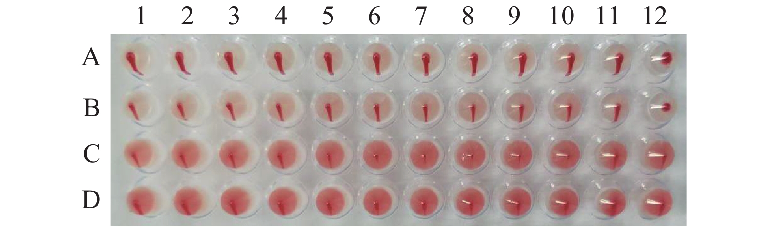
图 2 分离病毒的血凝性检测
注: 1~10为病毒滤液从1/2到1/1024梯度稀释;11,PBS空白对照;12,H9N2阳性对照;A,鸭血液;B,鸡血液;C,小鼠血液;D,兔血液。
Figure 2. Hemagglutination detection on isolated viruses
Note: 1–10: Virus filtrates diluted at a gradient of 1/2 to 1/1 024; 11: PBS blank control; 12: H9N2 positive control; A: duck blood; B: chicken blood; C: mouse blood; D: rabbit blood.

图 3 动物感染及其剖检特征
注:A和B为注射生理盐水的鸭和肝脏;C和D为注射病料研磨液的鸭和肝脏。
Figure 3. Infection and dissection on animals
Note: A: Duck injected with saline; B: liver of duck injected with saline; C: duck injected with ground liquid containing diseased material; D: liver of duck injected with ground liquid containing diseased material.
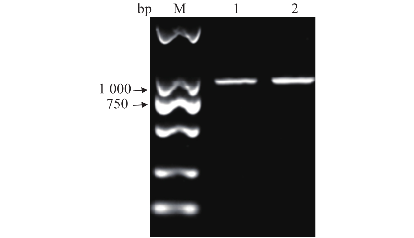
图 4 DHV-I的PCR鉴定
注:M,Marker DL2000;1,DHV-I标准株的扩增片段;2,Fujian2015的扩增片段。
Figure 4. PCR identification on DHV-I
Note: M: Marker DL2000; 1: amplified fragment of DHV-I standard strain; 2: amplified fragment of Fujian 2015.
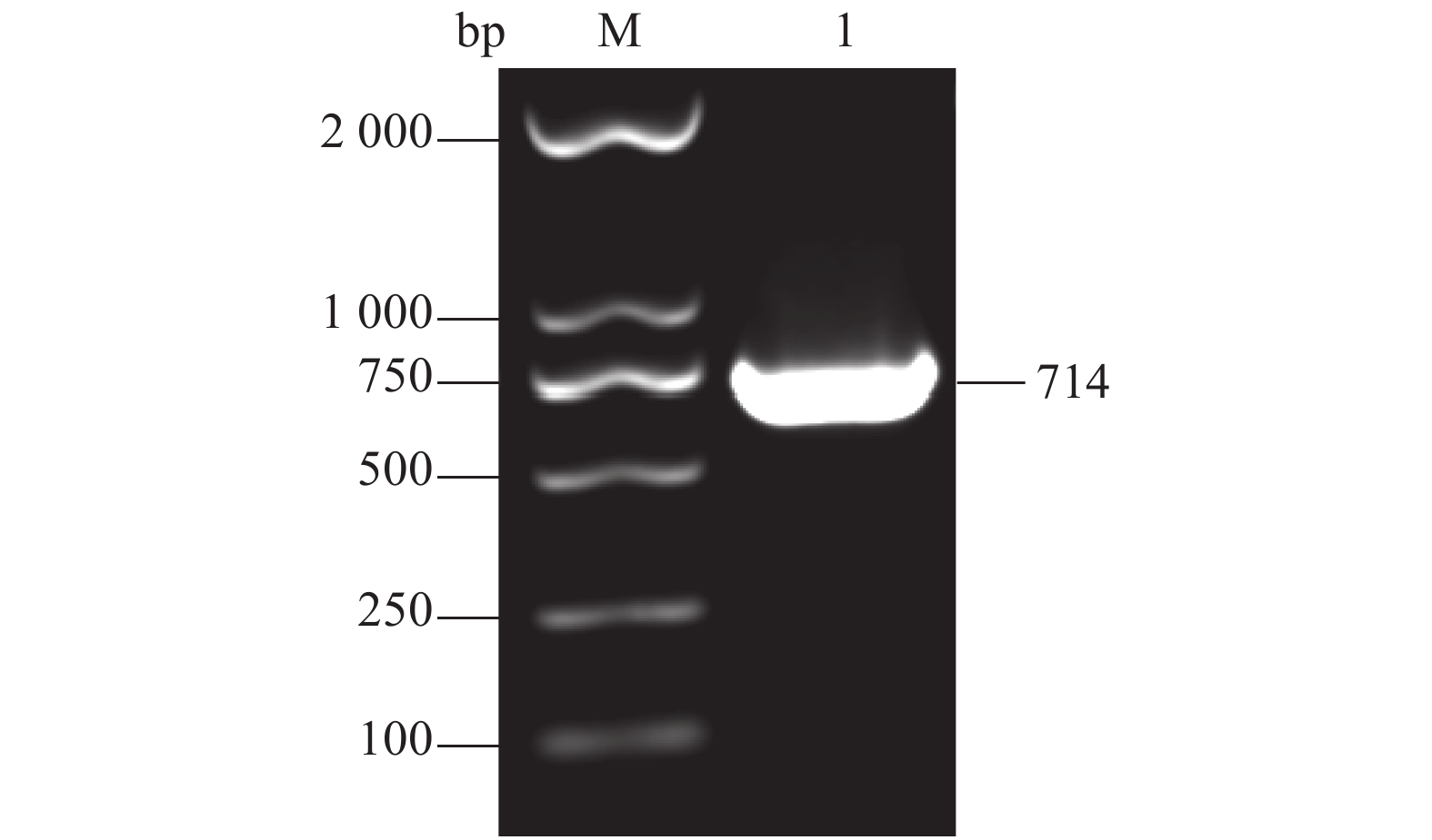
图 7 VP1基因PCR扩增
注:M,Marker DL2000;1,VP1的扩增片段。
Figure 7. PCR amplification of VP1
Note: M: Marker DL2000; 1: amplified fragment of VP1.
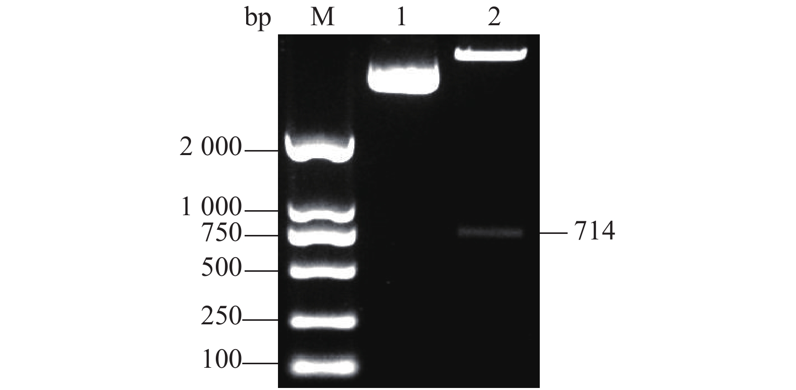
图 8 重组质粒pGEX-4T-1-VP1的酶切鉴定
注:M. Marker DL2000;1. 重组质粒pGEX-4T-1-VP1;2. 重组质粒pGEX-4T-1-VP1双酶切。
Figure 8. Restriction digestion on recombinant plasmid pGEX-4T-1-VP1
Note: M: Marker DL2000; 1: recombinant plasmid pGEX-4T-1-VP1; 2: double digestion of recombinant plasmid pGEX-4T-1-VP1.
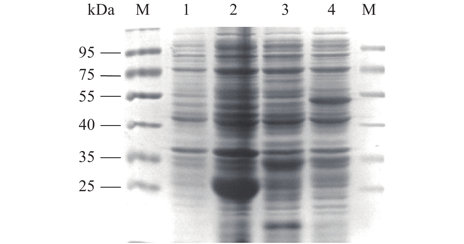
图 9 融合蛋白GST-VP1的SDS-PAGE分析
注:M为蛋白Marker; 1为质粒pGEX-4T-1诱导前表达产物; 2为质粒pGEX-4T-1诱导后的表达产物; 3为 质粒pGEX-4T-1-VP1诱导前的表达产物;4为质粒pGEX-4T-1-VP1诱导后的表达产物。
Figure 9. Fusion protein GST-VP1 by SDS-PAGE
Note: M: Protein marker; 1: plasmid pGEX-4T-1 expression product before induction; 2: expression product induced by plasmid pGEX-4T-1; 3: plasmid pGEX-4T-1-VP1 expression product before induction; 4: expression product induced by plasmid pGEX-4T-1-VP1.
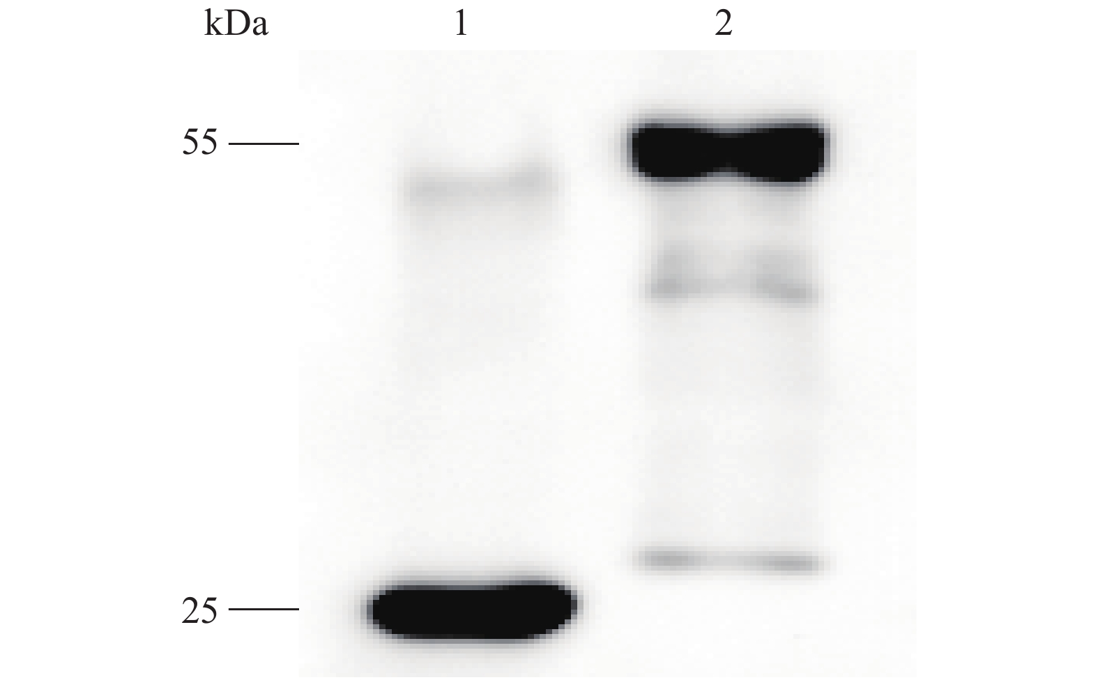
图 10 融合蛋白GST-VP1的Western-blotting分析
注:1,pGEX-4T-1诱导后的表达产物;2,pGEX-4T-1-VP1诱导后的表达产物。
Figure 10. Western-blotting on fusion protein GST-VP1
Note: 1: Expression product induced by plasmid pGEX-4T-1; 2: expression product induced by plasmid pGEX-4T-VP1.
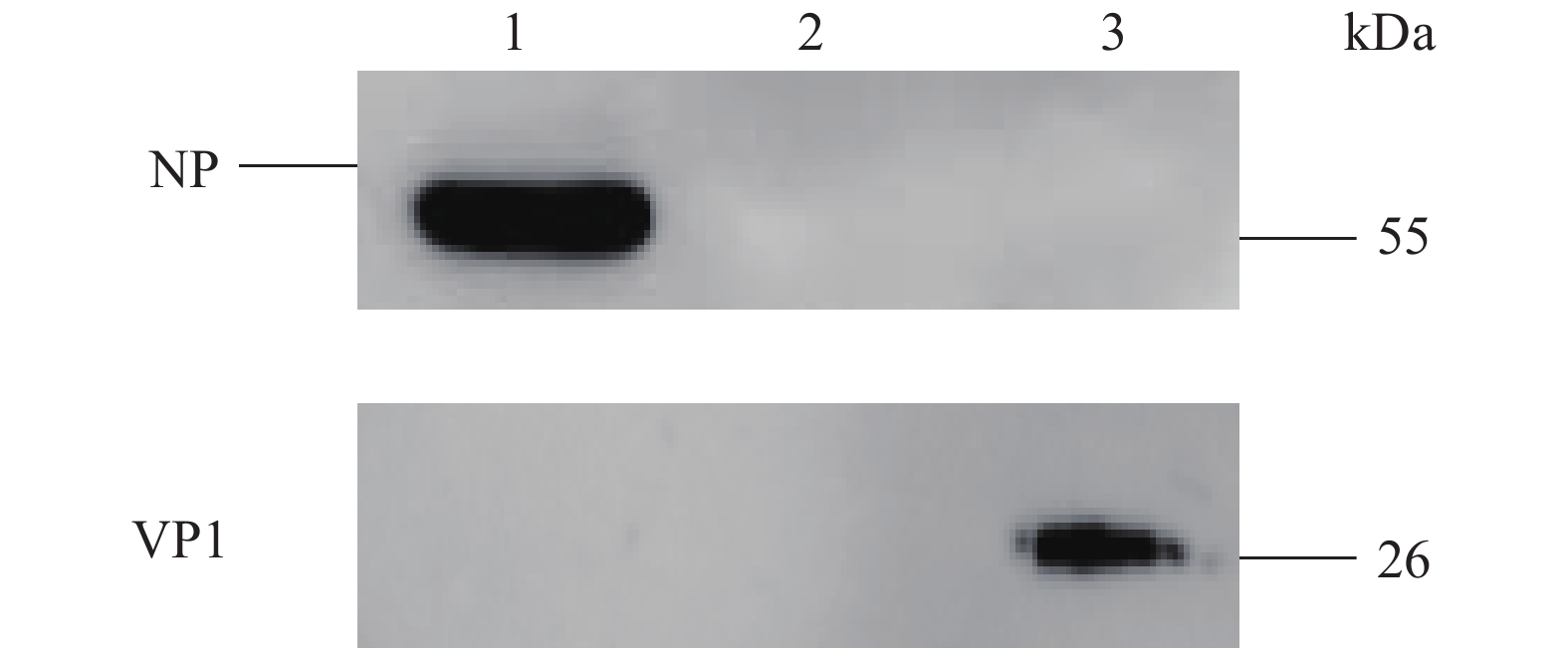
图 11 VP1多抗血清的Western-blotting分析
注:1:H9N2感染的A549细胞;2:健康鸭的肝脏;3:DHV感染的鸭肝脏。
Figure 11. Western-blotting on poly-antiserum against VP1
Note: 1: A549 cells infected with H9N2; 2: liver of control duck; 3: liver of DHV-infected duck.
表 1 分离的病毒与部分DHV毒株的VP1氨基酸序列比对
Table 1. VP1 amino acid sequences of isolated and partial DHV viruses
毒株
VirusVP1突变位点
VP1 mutation site256 aa 334 aa 547 aa Fujian2015毒株
Fujian2015 virusT M G 部分DHV毒株
Part of DHV virusesL/S V/T A/T  下载: 导出CSV
-
[1] KIM M C, KWON Y K, JOH S J, et al. Differential diagnosis between type-specific duck hepatitis virus type 1 (DHV-1) and recent Korean DHV-1-like isolates using a multiplex polymerase chain reaction [J]. Avian Pathology, 2008, 37(2): 171−177. doi: 10.1080/03079450801918670 [2] 何淼, 张宝康, 粟灵琳, 等. 磷酸化修饰对山豆根多糖抗Ⅰ型鸭肝炎病毒效果的影响 [J]. 南京农业大学学报, 2021, 44(2):322−330. doi: 10.7685/jnau.202003030HE M, ZHANG B K, SU L L, et al. Effect of phosphorylation on resisting duck hepatitis virusⅠ effect of Bush Sophora Root polysaccharide [J]. Journal of Nanjing Agricultural University, 2021, 44(2): 322−330.(in Chinese) doi: 10.7685/jnau.202003030 [3] 王宇飞, 孙伟, 陈佶慧, 等. 两株1型鸭甲肝病毒山东分离株的鉴定与致病性分析 [J]. 中国动物传染病学报, 2021, 29(1):26−30.WANG Y F, SUN W, CHEN J H, et al. Identification and pathogenicity of two duck hepatitis A virus type 1 isolated in Shandong, China [J]. Chinese Journal of Animal Infectious Diseases, 2021, 29(1): 26−30.(in Chinese) [4] 李志辉. 南方地区鸭病毒性肝炎的诊治 [J]. 当代畜禽养殖业, 2019(2):30. doi: 10.3969/j.issn.1005-5959.2019.02.025LI Z H. Diagnosis and treatment of duck viral hepatitis in southern China [J]. Modern Animal Husbandry, 2019(2): 30.(in Chinese) doi: 10.3969/j.issn.1005-5959.2019.02.025 [5] NIU Y J, MA H Y, DING Y H, et al. The pathogenicity of duck hepatitis A virus types 1 and 3 on ducklings [J]. Poultry Science, 2019, 98(12): 6333−6339. doi: 10.3382/ps/pez455 [6] 韩宜均, 孙举, 原昆鹏, 等. 抗Ⅰ型鸭肝炎病毒单链抗体库的构建与筛选 [J]. 黑龙江畜牧兽医, 2021(3):91−96.HAN Y J, SUN J, YUAN K P, et al. Construction and screening of ScFv library against Duck hepatitis virus type Ⅰ [J]. Heilongjiang Animal Science and Veterinary Medicine, 2021(3): 91−96.(in Chinese) [7] WANG A P, GU L L, WU S, et al. Duck hepatitis A virus structural proteins expressed in insect cells self-assemble into virus-like particles with strong immunogenicity in ducklings [J]. Veterinary Microbiology, 2018, 215: 23−28. doi: 10.1016/j.vetmic.2017.12.020 [8] 毛赛. 1型鸭肝炎强毒对成鸭致病、体内分布及免疫发生特性研究[D]. 雅安: 四川农业大学, 2019MAO S. Study on the distribution, pathogenicity and immunogenesis of adult ducks infected with virulent duck hepatitis A virus 1[D]. Yaan: Sichuan Agricultural University, 2019. (in Chinese) [9] WANG A P, LIU L, GU L L, et al. Expression of duck hepatitis A virus type 1 VP3 protein mediated by avian adeno-associated virus and its immunogenicity in ducklings [J]. Acta Virologica, 2019, 63(1): 53−59. doi: 10.4149/av_2019_104 [10] LAI Y L, ZENG N, WANG M S, et al. The VP3 protein of duck hepatitis A virus mediates host cell adsorption and apoptosis [J]. Scientific Reports, 2019, 9(1): 16783. doi: 10.1038/s41598-019-53285-0 [11] WEN X J, CHENG A C, WANG M S. Sequence analysis and B cell epitope prediction of duck hepatitis A virus 1 VP1 gene [J]. Advanced Materials Research, 2013, 647: 214−219. doi: 10.4028/www.scientific.net/AMR.647.214 [12] 王平, 潘文石, 胡寿文, 等. 北京小鸭病毒性肝炎的研究:(一)诊断和防治 [J]. 北京大学学报(自然科学版), 1980, 16(1):55−74.WANG P, PAN W S, HU S W, et al. Duck virus hepatitis in Peking duckling I. Diagnosis and prevention [J]. Acta Scicentiarum Naturalum Universitis Pekinesis, 1980, 16(1): 55−74.(in Chinese) [13] XUE W X, ZHAO Q, LI P F, et al. Identification and characterization of a novel nanobody against duck hepatitis A virus type 1 [J]. Virology, 2019, 528: 101−109. doi: 10.1016/j.virol.2018.12.013 [14] 张丽. 河北省鸭肝炎病毒BD-Ⅰ株的分离鉴定及弱毒株培育[D]. 保定: 河北农业大学, 2008.ZHANG L. Isolation, identification and cultivation of duck hepatitis virus BD-I strain in Hebei Province. [D]. Baoding: Agricultural University of Hebei, 2008. (in Chinese) [15] 杜君. 鸭病毒性肝炎诊断与防治 [J]. 畜牧兽医科学, 2020(13):139−140.DU J. Diagnosis and control of duck hepatitis [J]. Graziery Veterinary Sciences, 2020(13): 139−140.(in Chinese) [16] 管飘萍. 2017~2019年山东省部分地区鸭甲肝病毒的分子特征分析和VP1基因的表达[D]. 扬州: 扬州大学, 2020.GUAN P P. Molecular characteristics of duck hepatitis A virus in some areas of Shandong Province from 2017 to 2019 and expression of VP1 gene[D]. Yangzhou: Yangzhou University, 2020. (in Chinese) [17] 傅秋玲, 傅光华, 陈红梅, 等. 胰腺炎型鸭1型甲肝病毒结构蛋白VP1基因的克隆和表达 [J]. 福建农业学报, 2014, 29(5):409−412. doi: 10.3969/j.issn.1008-0384.2014.05.001FU Q L, FU G H, CHEN H M, et al. Cloning and prokaryotic expression of the VP1 gene of pancreotropic duck hepatitis type 1 virus [J]. Fujian Journal of Agricultural Sciences, 2014, 29(5): 409−412.(in Chinese) doi: 10.3969/j.issn.1008-0384.2014.05.001 [18] 王莹, 张兴晓, 朱洪伟. 鸭甲型肝炎病毒VP1研究进展 [J]. 病毒学报, 2021, 37(5):1260−1267.WANG Y, ZHANG X X, ZHU H W. Research progress on VP1 of duck hepatitis A virus [J]. Chinese Journal of Virology, 2021, 37(5): 1260−1267.(in Chinese) -


 点击查看大图
计量
- 文章访问数: 445
- HTML全文浏览量: 148
- PDF下载量: 19
- 被引次数: 0
